
Blood flow assessment
Doppler imaging shows blood flow in and around breast tissue or lumps, helping us interpret findings.
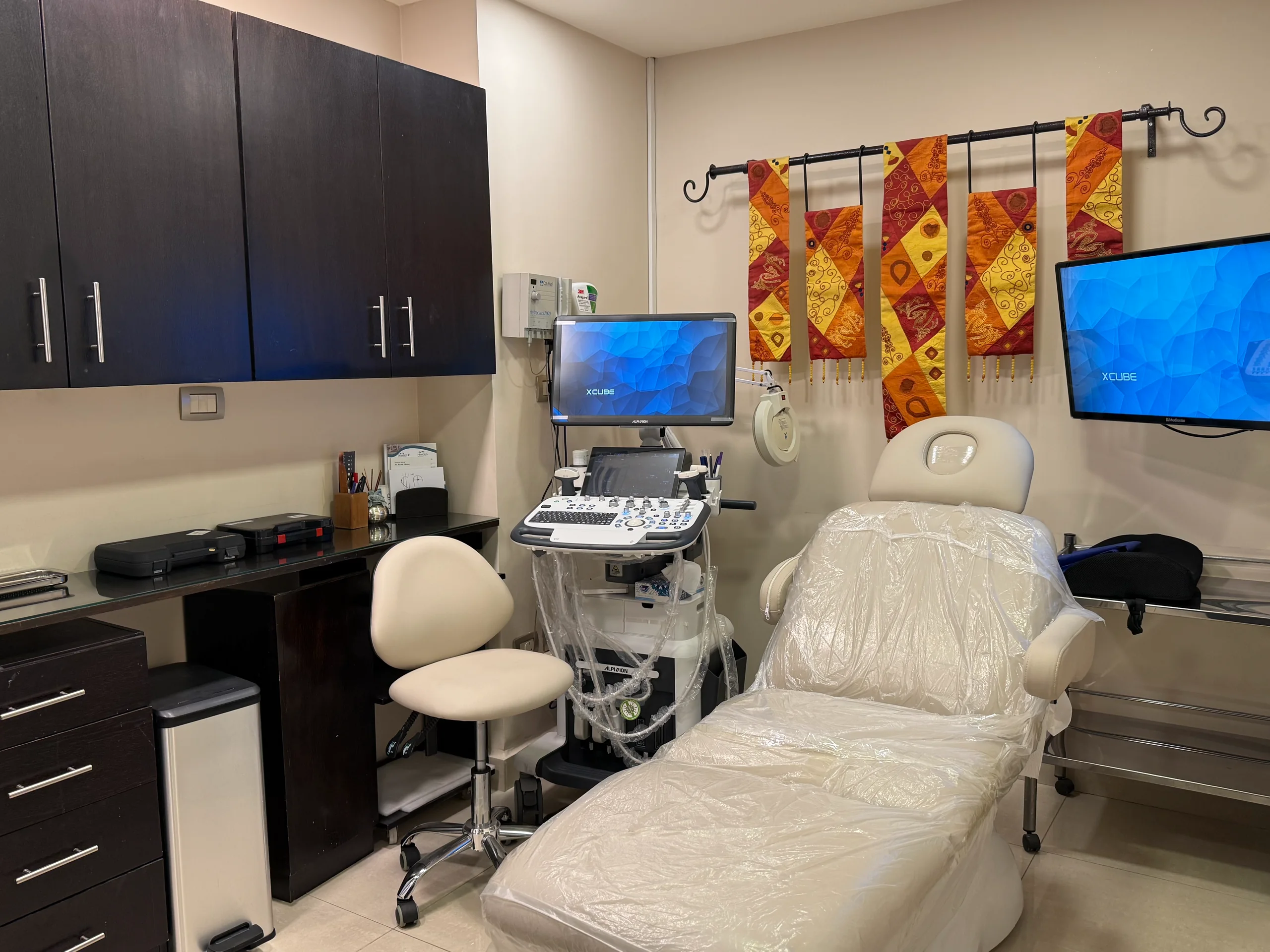
At the clinic, high-frequency ultrasound helps us look closely at your breast tissue, assess changes, lumps, cysts or fluid collections, and examine lymph nodes in the underarm when clinically needed.
Depending on your needs, imaging may include these techniques:
Doppler imaging shows blood flow in and around breast tissue or lumps, helping us interpret findings.
Elastography can help assess tissue stiffness and lump characteristics alongside conventional ultrasound.
Ultrasound helps assess the location, size, shape and borders of a lump and whether it's solid or fluid-filled. We then discuss if further tests are needed.
When a breast tissue sample is needed, ultrasound guidance helps your doctor target the area accurately. Samples are taken using sterile techniques and appropriate medical safety precautions.
If breast pain keeps affecting your daily life, we look at possible causes and plan care around your findings. Specialist treatments or procedures may be considered when medically appropriate.
A closer look at breast and chest pain
High-frequency ultrasound can help us assess certain causes of breast or chest pain. It allows us to examine your breast tissue, look for changes and check for lumps, cysts or fluid collections when needed.
Other imaging techniques help assess breast tissue structure, elasticity and blood flow, adding useful information about changes found during your examination.
Imaging helps us assess a lump or unusual area and decide whether further testing or a tissue sample is needed.
A full assessment may avoid unnecessary procedures. When a biopsy is needed for an accurate diagnosis, we explain why it matters.
Ultrasound helps us examine your breast's structure, including glandular and fatty tissue, and look for changes that may need further review.
We review results alongside your age, health and medical history. Mammography may also be recommended to assess breast density.
When clinically needed, a specialist gynecological assessment may include an ultrasound to measure the thickness of your uterine lining.
Ultrasound can help assess chest wall muscles, including their thickness, structure and any relevant changes.
When symptoms suggest nerve involvement, specialist tests such as nerve conduction studies may help us assess nerve function and whether it could contribute to your pain.